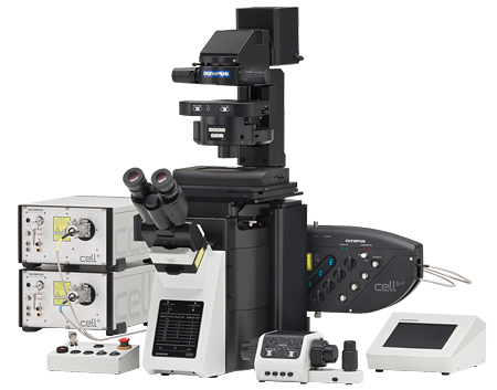
Higher Level of Live Cell Research
Deck 시스템 도입으로 뛰어난 확장성
장시간의 이미징 촬영에도 최적의 안정성제공
최상의 광학성능 제공
터치패널과 소프트웨어를 통한 완벽제어가능
사용자의 편의를 위한 인체공학 디자인
2개의 “Deck”구조를 채용 다양한 이미지기술에 필요한 여러 장치들을 장착 할 수 있게 되었습니다.
또한 오랜 시간 동안 세포관찰에 용이 할 수 있도록 높은 내구성과 안정성을 가지고 있어 장비의 활용성을 극대화 시킬 수 있습니다.
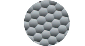
● Fly-eye를 적용 sCMOS와 같이 큰 영상획득장치에서 균일한 영상을 획득할 수 있습니다.
● Ultrasonic 전자동 제물대를 채용 진동과 소음 없이 정확한 이동이 가능하게 되었습니다.
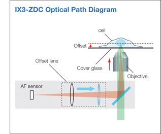
● 새로운 Zero-Drift Compensation를 장착 짧은시간, 오랜시간동안 포커스 변화 없는 영상을 획득 할 수 있습니다.
● 새롭게 개발된 고속 Shutter 및 Filter Wheel을 통해 정확한 이미지 획득이 가능하게 되었습니다.
● Real Time Controller를 사용하여 빠른 세포변화에 대응할 수 있도록 설계 되었습니다.
개선된 광학성능
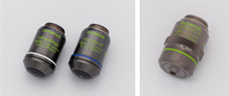
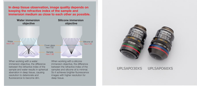
● 고해상도의 PH영상과 형광을 렌즈변경없이 획득 가능한 새로운 렌즈 적용
● iPS/ES와 같은 floating cell에 특화된 새로운 렌즈 적용
● Fly-Eye 적용으로 밝고 균일한 조명을 쉽게 얻을 수 있음
라이브 셀을 위한 최고의 선택
One shot모드를 통한 장시간의 세포관찰, Continuous 모드를 사용 TIRF 및 Calcium 이온관찰 시 빠른 세포의 변화를 포커스 변화 없이 관찰 가능. IR레이저 사용을 Low phototoxicity 제공.
기존의 제품에 비해 정밀도, 정확도가 개선됨.
ZDC-Continuous mode
Evanescent field : 200nm
Lens: APON60XOTIRFMAPO 60X, NA 1.49, WD 0.1mm, Correction collar
Laser: 491nm 50mW
Camera: Andor iXon-888
시료의 굴정률(1.38)과 가까운 1.4의 굴절률을 사용하여 정확한 3차원영상관찰 및 장시간의 세포관찰에도 보다 선명한 영상 구현 가능
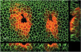
Enlarged view shows invaginating tracheal placode.
사용자 편의개선

렌즈의 배율에 맞게 밝기를 자동 보정하는 기능 제공
● 퀠러 조절이 편안한 디자인
앞쪽으로 변경된 콘덴서 조절 레버와 스톱퍼를 통한 콘덴서 오작동을 방지함.
● 현미경 오염방지 기술 적용
현미경 하단에 오염 방지 막을 사용 이물질에 의해 S/N 저하를 방지
● Touch Panel - 터치 패널을 통한 현미경의 모든 제어가 가능, ZDC기능도 터치패널을 통해 제어가 가능
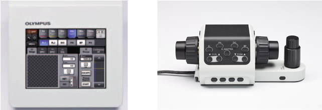
렌즈변환, 필터휠, 셔터, 스테이지 이동 사용자 편의를 배려한 수동조절 콘트롤러
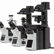
높은 확장성과 이미징을 위한 향상된 정확성
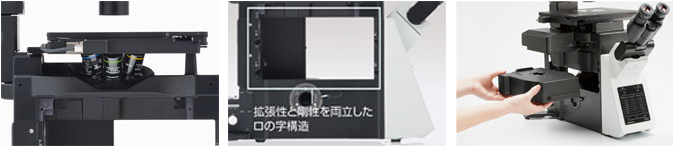
노즈피스와 가깝게 Z축 구성 4각형의 구조로 증가된 안정성 손쉬운 Modify 가능
● 고속의 Filter wheel, shutter 적용
- 60ms의 빠른 필터휠과 27ms의 속도로 개폐가 가능한 셔터를 적용 고속의 영상 촬영에 최적화 되었음.
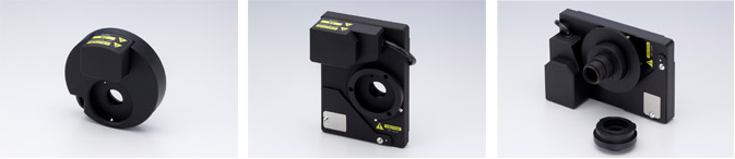
27MS의 고속셔터 60MS의 고속 Excitation 필터 휠 60ms의 고속 Emission 필터 휠
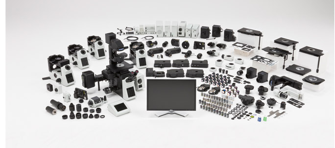
● 수많은 유저들의 요구를 충족할 수 있도록 다양한 전동 옵션 유닛들로 구성되어 있습니다.
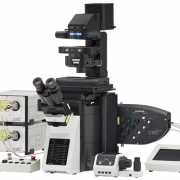
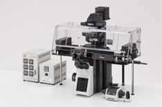
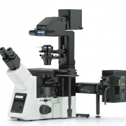
Live Cell System (IX83-ZDC)
“Continuous Auto-Focus Function for Live Cell Imaging” - 손쉬운 자동화 시스템 |
|
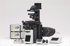
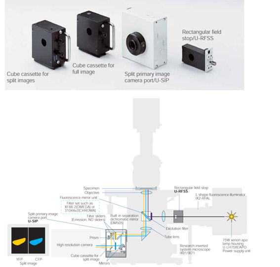
TIRF
“Perfect TIRF Imaging” - 레이저별 Evanescence field 보정 색수차 없이 수 없는 영상 제공 |
|
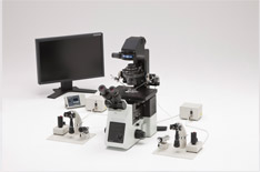
ON3
“From Microinjection to patch clamping” - 조이스틱을 이용한 손쉬운 조작 |
|
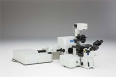
FV1000
“From Imaging to Analysis” - 고속, 고해상도의 Confocal 이미지 촬영 |
|
FV1000MPE
“Advanced Deeper Imaging with High Resolution” - 1mm 정도의 두꺼운 샘플 3D 구현 가능 |
|
FRET System
- 장착이 쉽게 설계되어 추가 장착에 따른 불편을 최소화
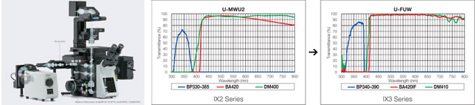
- 유사한 장치에 비해 10%높은 투과율을 가지고 있음
- 소프트웨어를 통한 정확한 FRET 분석가능
Specifications
Microscope Frame |
Optical System |
UIS2 Optical System |
|---|---|---|
|
Revolving nosepiece |
Motorized sextuple revolving nosepiece (DIC slider attachable), simple waterproof structure. |
|
|
Focus |
Stroke: 10.5 mm Minimum increment: 0.01μm, Maximum nosepiece movement speed: 3 mm/s |
|
|
Light Path selection |
Motorized 0:100/50:50/100:0 (Left side port: BI port) |
|
|
Transmitted light illuminator |
Pillar tilt mechanism (30° inclination angle, with vibration reducing mechanism), Condenser holder (with 88 stroke, refocusing mechanism), Field iris diaphragm adjustable, 4 filter holders Light source: * 12 V 100 W halogen bulb (pre-centered) * High color reproductive LED light source |
|
|
Observation tube |
Widefield (FN 22) |
*Widefield tilting binocular * Widefield binocular * Widefield trinocular |
|
Stage |
Scanning stage with ultrasonic |
Stage stroke: X:76 mm x Y: 52 mm, maximum stage movement speed: 30 mm/s |
|
Mechanical stage with right handle |
||
|
Right handle stage |
||
|
Flexible right handle stage |
||
|
Gliding Stage |
||
|
Plain Stage |
||
|
Condenser |
Motorized long working distance condenser |
W.D. 27 mm, NA 0.55, motorized turret with 7 position slots for optical devices (3 positions for ø30 mm and 4 positions for ø38 mm), motorized aperture and polarizer. |
|
Long working distance universal condenser |
||
|
Long working distance relief contrast |
||
|
Ultra long working distance |
||
|
Fluorescence Illuminator |
L-shape-fluorescence illuminator with flyeye lens |
L-shaped design with exchangeable FS module. |
|
L-shape-fluorescence illuminator |
L-shaped design with exchangeable FS and AS modules. | |
|
Fluorescence illuminator |
Straight design with field iris diaphragm. | |
|
Fluorescence Mirror Turret |
Motorized fluorescence mirror turret |
Motorized turret with 8 positions, built -in shutter, simple waterproof structurer. |
|
Coded fluorescence mirror |
N/A | |
|
Fluorescence mirror turret |
N/A |
|
|
Fluorescence light source |
*130W Hg light guide illumination * 100W Hg apo lamp housing and transformer *100W Hg lamp housing and transformer * 75W Xe lamp housing and transformer. |
|
|
Focus Compensator |
Z-drift Compensator |
offset method (Focus search, one-shot focus, continuous focus) |
|
Class 1 laser device |
||
|
Filter wheel/shutter |
Motorized fast filter wheel |
High speed mode 60 ms, low vibration mode 100 ms (rotation time until next hold on the wheel) |
|
Motorized fast filter wheel for emission |
High speed mode 60 ms, low vibration mode 100 ms (rotation time until next hold on the wheel) C-mount adapter and bayonet mount adapter are enclosed. |
|
|
Motorized fast shutter |
High speed mode 26.2 ms, Low vibration mode 60 ms (rotation time on one way). |
|
|
Motorized attenuator wheel |
Time to shift another filter 300ms (rotation time until next hole on the wheel). |
|
|
Operating environment |
*Indoor use *Ambient temperature: 5° to 40° C (41° to 104° F) *Maximum relative humidity: 80% for temperatures up to 31°C (88°F), decreasing linearly through 70% at 34°C(93°F), 60% at 37°C (99°F), to 50% relative humidity at 40°C (104°F) *Supply voltage fluctuations: Not to exceed | |
Catalog download : IX83.pdf
 IX73
IX73